Description
Sub-4 Technologies are delighted to introduce an innovative concept in clinical biomechanics, which is considered by many to be an important part of the future of musculoskeletal (MSK) health care.
The Digital Pelvic Inclinometer (DPI) allows the practitioner to understand and measure the role of the pelvis in development of repetitive injury including lower back.
What is a DPI? Explaining the features of this new innovation.
Video 1.
Video 1. explains what a Digital Pelvic Inclinometer (DPI) is, and its features.
How to hold a DPI and where to place it on the pelvis.
Video 2.
Video 2. show you how to hold the DPI and where to place it on the patient’s pelvis to quantify position.
How to quantify pelvic torsion using the DPI.
Video 3.
Video 3. explains how to quantify pelvic torsion using a simple clinical protocol.
What does it do?
Using a DPI allows the practitioner to expand their clinical biomechanics skills to a much higher level, opening up a whole new realm of MSK understanding.
- The DPI is an easy to use hand-help instrument designed to allow the practitioner to measure innominate inclincation and establish pelvic torsion.
- The two moveable arms with sensory finger grips increase practitioner proprioception and therefore accurate measuring.
- The precision arms pivot about a main body, which houses a digital inclinometer and a spirit level.
- The DPI allows the practitioner to record quantitive evidence of MSK dysfunction and therefore evidence of correction after treatment.
What are the clinical applications?
DPI in clinical use measuring pelvic torsion
The DPI is easy to use clinically, giving the patient confidence that the practitioner is a specialist at the cutting edge of technology.
Following DPI clinical protocol allows the practitioner to establish whether a leg length inequality is anatomical, apparent or functional.
The numerical reading is said to be +ve (positive) if the ASIS is lower than the PSIS during measuring, from a horizontal reference plane. The reading is said to be -ve (negative) is the ASIS is higher than the PSIS, from the same reference plane. Clinically normal innominate inclinication has been reported between 8 to 10 +ve, although different body types and ages can deviate these ranges (1,2). By understanding the behaviour of the innominates and sacrum, the practitioner is able to establish the difference between anatomical, apparent and functional leg length inequality; demonstate correction of the pelvic torsion and understand the role of the pelvic mechanics in the kinetic chain witout the need for a scan.
How does it work?
Data is captured by the DPI via a tiny three-axis accelerometer attached to a small electronics board. The electronics use this data to calculate the angle of tilt, This data is shown in numerical form on the liquid crystal display (LCD).
How is the DPI held?
The practitioner places the index finger and thumb on each hand on each finger grip at the end of the DPI arms. With each index finger slightly prominent ready for concurrent palpation of the posterior superior iliac spine (PSIS) and anterior superior iliac spine (ASIS), the practitioner positions the DPI on the side of the innominate bone and takes a reading. The practitioner moves their index finger over the most prominent point of the iliac crests until the apex is established for the measuring. The practitioner then reads off the degree of inclinication from the LCD. The difference in inclination between each innominate established the degree of pelvic torsion.
What will I get for my investment?
A Digital Pelvic Inclinometer.
One full-days training with Clifton Bradeley and patient.
An Operator’s Manual including clinical case studies.
A 2 year guarantee.
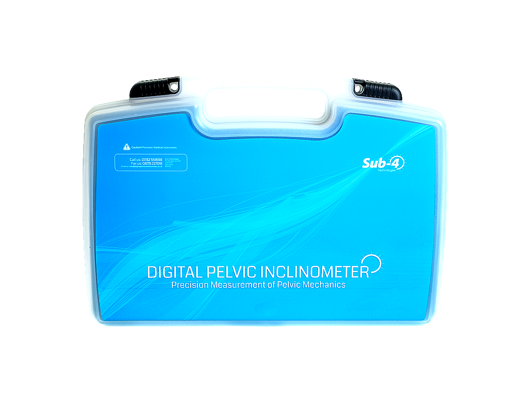
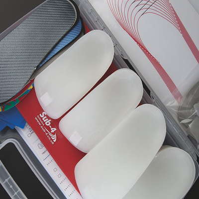
A protective case.
On-going support
Practitioners who purchase a DPI will have access to on-going support and training from Sub-4 Technologies.
Users will be invited to attend advanced training courses in the use of the DPI and how to work within the realm of pelvic mechanics.
Training is carried our at Sub-4 Health in Staffordshire.
References
1. Levine, D. and M.Whittle (1996). “The Effects of Pelvic Movement on Lumbar Lordosis in the Standing Position.
“Journal of Orthopaedic & Sports Physical Therapy. 24(3): 130 – 135
2. Cummings G, S.J., Barnes K. (1993). “The Effects of Imposed Leg Length Difference on Pelvic Bony Symmetry.
“Spine 18(3): 368 – 373


